Margo Case Study Analysis
Upon examining the results of the MRI scan of her brain, Margo noted that there were visible and notable differences between her brain’s MRI results and those of a normal brain. She noticed that the cortex of her brain’s frontal lobe had a shrinkage that exceeded the expected shrinkage related to age. There lacked a clear differentiation between the brain’s white and gray matter. Her lateral ventricles are hypointense and partially dilated. All of these changes exceed the age-related changes of the brain with reference to her age. Normal age-related cortical atrophy is usually observed in adults between 50 and 65 years (Cheng et al., 2020).
Margo has complained of problems with her thinking and focus. These problems are related to the extensive shrinkage of the frontal lobe. Notably, the frontal lobes are associated with the control of cognitive functions and memory creation and maintenance (Stuss & Benson, 2019). This means that the shrinking of the frontal lobe has an impact on her intellectual capabilities, including her ability to think and focus. The frontal lobe shrinkage also affects brain structures, such as the cerebellum, associated with the coordination of body parts and balance. The reduced differentiation of the white, as indicated in the MRI scan images, can indicate a developing cerebral abscess, which can affect blood flow, leading to confusion, body weakness, and blurred vision, which may affect movement and focus.
If Margo continues drinking alcohol, she will experience progressive frontal atrophy, white matter damage, and alcoholic cerebellar degeneration. Margo will be at a high risk of increased memory loss and dementia symptoms, alcoholic myopathy, body tremors, inability to maintain balance or walk for a distance, falls, hallucinations, and confusion.
References
Cheng, Y., Xu, J., Dong, C., Shen, Z., Zhou, C., Li, N., … & Xu, X. (2020). Age-related atrophy of cortical thickness and genetic effect of ANK3 gene in first-episode MDD patients. NeuroImage: Clinical, 28, 102384. https://doi.org/10.1016/j.nicl.2020.102384
Stuss, D. T., & Benson, D. F. (2019). The frontal lobes control of cognition and memory. In The frontal lobes revisited (pp. 141-158). Psychology Press.
ORDER A PLAGIARISM-FREE PAPER HERE
We’ll write everything from scratch
Question 
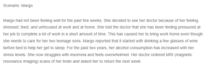
Scenario: Margo
Margo had not been feeling well for the past few weeks. She decided to see her doctor because of her feeling stressed, tired, and unfocused at work and at home. She told the doctor that she has been feeling pressured at her job to complete a lot of work in a short amount of time. This has caused her to bring work home even though she needs to care for her two teenage sons. Margo reported that it started with drinking a few glasses of wine before bed to help her get to sleep. For the past two years, her alcohol consumption has increased with her stress levels. She now struggles with insomnia and feels overwhelmed. Her doctor ordered MRI (magnetic resonance imaging) scans of her brain and asked her to return the next week.
Margo Case Study Analysis
Margo returned to see her doctor a week later. During that appointment, the doctor showed her the brain scan and explained that she should begin to make some lifestyle changes. She was struck by the differences between her brain and a healthy one and promised to make some changes.
https://purdueglobal.brightspace.com/content/enforced/220321-042-2203B-PS215-01-2475962/Picture1.png?_&d2lSessionVal=l8zbEUTWBBk0xHbNSKlLMrJH9
Brain scan showing alcoholic brain versus normal brain
Using the scenario above, explain what Margo saw in the MRI scan of her brain. Why is she likely suffering from problems with her thinking and focus? Make some predictions about what kinds of neurological problems she will develop if she continues to drink.
Textbook:
Ward, J. (2019). The Student’s Guide to Cognitive Neuroscience (4th Edition). Taylor & Francis. https://purdueuniversityglobal.vitalsource.com/books/9781351035163