Hemolysis
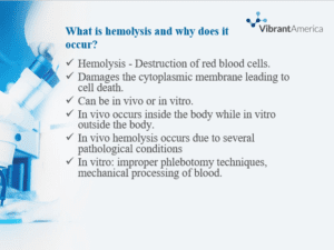
Hemolysis is the breakdown of erythrocytes, releasing their contents into the blood plasma (Rifai, 2017). It can occur in vivo or in vitro. Hemolysis damages the cytoplasmic membrane of erythrocytes, resulting in lysis and cell death (Rifai, 2017). In vivo, hemolysis can occur due to many pathological conditions. They include bacteria such as streptococcus, parasites such as malaria, autoimmune conditions, and genetic disorders such as sickle cell disease (Rifai, 2017). In vitro, hemolysis can occur due to the effects of mechanical blood processing during surgery, such as centrifuging, improper technique during blood specimen collection, and actions of bacteria in cultured blood specimens (Wan Azman et al., 2019).
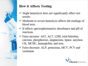
Slight hemolysis has no significant effect on most laboratory rest values, but moderate to severe hemolysis will directly affect spectrophotometric absorbance readings and alter the pH readings. Aspartate aminotransferase (AST) and lactate dehydrogenase (LDH) have higher activity with the red blood cells compared with plasma, and hence, their activities will be enhanced in hemolysis (Makowski, 2020). Hemolysis may falsely increase AST and alanine transaminase (ALT). LDH, total bilirubin, phosphorous, calcium, magnesium, lipase, amylase, creatine kinase (CK), hemoglobin, iron, and mean corpuscular hemoglobin concentration (MCHC).On the other side, it may falsely decrease alkaline phosphatase (ALP), creatinine, packed cell volume (PCV), potassium, and mean corpuscular volume (MCV) (Makowski, 2020).
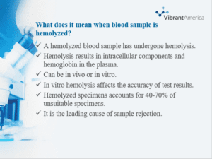
A hemolysed blood sample has undergone hemolysis. Hemolysed specimens have hemoglobin and intracellular components of the red blood cells and hemoglobin in the extracellular space of blood (Wan Azman et al., 2019). The hemolysis can be in vivo or in vitro. In vitro, hemolysis is undesirable since it affects the dependability of laboratory testing and the accuracy of the results. It can occur due to mishandled or improper procedures during specimen collection (Wan Azman et al., 2019). Hemolysed specimens account for 40-70% of all unsuitable samples identified (Wan Azman et al., 2019). In vitro, hemolysis is the leading cause of sample rejection in inpatient and outpatient samples (Wan Azman et al., 2019).
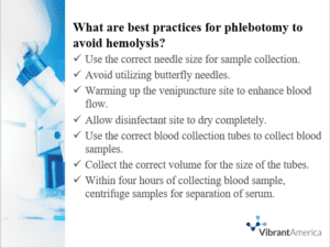
Prevention of hemolysis is important to preserve the quality of a serum sample for testing. The best practices to prevent hemolysis include:
- Use the correct needle size for sample collection.
- Avoid utilizing butterfly needles.
- Warming up the venipuncture site to enhance blood flow.
- Allow the disinfectant site to dry completely.
- Use the correct blood collection tubes to collect blood samples.
- Collect the correct volume for the size of the tubes.
- Within four hours of collecting a blood sample, centrifuge samples for the separation of serum (CDC, 2021).
CDC. (2021, June 11). A quick-reference tool for Hemolysis status. Centers for Disease Control and Prevention. https://www.cdc.gov/ncezid/dvbd/stories/research-lab-diagnostics/hemolysis-palette.html
Makowski, G. (2020). Advances in clinical chemistry (1st ed.). Academic Press.
Rifai, N. (2017). Tietz textbook of clinical chemistry and molecular diagnostics – E-book. Elsevier Health Sciences.
Wan Azman, W. N., Omar, J., Koon, T. S., & Tuan Ismail, T. S. (2019). Hemolyzed specimens: Major challenge for identifying and rejecting specimens in clinical laboratories. Oman Medical Journal, 34(2), 94-98. https://doi.org/10.5001/omj.2019.19
ORDER A PLAGIARISM-FREE PAPER HERE
We’ll write everything from scratch
Question

Hemolysis
What is hemolysis, and why does it occur?
How it affects testing
What does it mean when a blood sample is hemolyzed?
What are the best practices for phlebotomy to avoid hemolysis?
Please use logo and logo colors.


