Forensic Anthropology and Odontology
Description of each of the identified bones above
The occipital bone is the back part of the skull and adjoins five other bones out of the seven that make up the skull. Its primary purpose is to enclose the back part of the brain and form a link between the spinal cord and the brain via the medulla oblongata.
Parietal bone – These are two bones on the skull’s right and left sides. They adjoin the frontal, occipital, temporal, and sphenoid bones. They mainly protect a large part of the brain on both sides.
Frontal Bone – This bone forms a large part of the skull. This bone underlies the forehead and extends backward to the coronal suture. It includes a joint with two small bones of the nose bridge and one of the cheekbones in the front. Its primary purpose is to protect the eyes and other forehead components.
Maxilla – Most of the facial skeleton is made up of the maxilla. This bone extends from the middle and lower part of the eye socket, forming the nose opening and nose aperture.
The mandible is the most prominent, lowest, and strongest bone of a human’s facial skeleton. It forms the lower jawline and holds the lower teeth. Additionally, this bone assists in mastication.
Clavicle – Also known as the collarbone, it is a slightly S-shaped bone that connects the sternum to the shoulder.
Scapula – Equally identified as the shoulder blade, it is a triangular bone on the upper back. This bone is part of a complex network of pieces that all coordinate to help in arm movements.
Sternum – Similarly known as the breastbone, it is a bone found in the middle of the chest. Its primary function is serving as a connection point for ribs, but it also helps protect organs in the chest region from injury.
Humerus – is the lengthy bone running from the shoulder to the elbow. It connects the scapula to the radius and ulna.
Ribs – are flat bones that form a protective cage for internal organs around the chest region. Humans have a total of 24 ribs in pairs of 12.
Vertebrae – These are a chain of interlocking bones, 33 in total, that runs from the base of the skull to the lower abdomen. Each vertebra serves three functions: transverse processes for alignment, load-bearing, and spinal cord protection.
Pelvic Girdles – Also known as the pelvis, are basin-shaped bones in the lower abdomen. They comprise the ilium, ischium, pubis, coccyx, and sacrum. Their main functions include supporting and containing lower abdomen organs, like intestines and reproductive organs, and connecting the core to the legs.
Radius and ulna – These two are the bones of the forearm. The radius is lateral bone, while the ulna is medial. Their primary function is pivoting around each other to allow wrist rotation.
Carpals, metacarpals, and phalanges are sets of hand bones, summing up to 27 bones. Carpals are a set of eight bones, metacarpals, i.e., fingers are five, and phalanges, i.e., bones of the fingers, are three for each finger except the thumb, which has two.
Femur – Likewise known as the thigh bone, it is the longest, strongest, and heaviest bone in the human body. Its primary function is the stability of gait and weight-bearing.
A patella is a small bone on the knee joint’s front side where the femur and tibia connect. Its main functions are helping bones glide easily, securing the upper and lower leg muscles, and protecting the knee joint.
Tibia – This is the larger of the two bones of the lower leg. Also known as the shinbone, its primary function is to carry most of the body weight.
Fibula – This is the lower leg’s smaller and much thinner bone. Its primary function is the attachment of muscles.
The talus, metatarsal bones, lateral cuneiform bones, and phalanges are the foot bones. The tarsals are seven, five metatarsals, and fourteen phalanges.
Part 2
Odontology and how it’s used in facial reconstruction
Simply put, odontology studies teeth, mainly their development, structure, and diseases that affect them. In forensic science, forensic odontologists are broadly trained that they can be able to assist in identifying unknown remains or tracing bite marks, among further areas of forensic science associated with teeth, as in this case, facial reconstruction, also known as a forensic facial approximation (FFA) (Chowdhry et al., 2018) 2016).
FFA is a two- or three-dimensional recreation of a person’s face from a skull’s remains, which gives an excellent semblance to the deceased person and helps identify them. FFA involves three stages; the first stage is anatomical modeling (Chowdhry et al., 2018). During anatomical modeling, odontologists must model the muscles and relevant structures on a skull, utilize strict anatomical guidelines of origin and insertion, and thoroughly know their existing variations.
The second stage is called morphology determination, which involves the morphology of soft tissue envelope over the skeletal covering in the crucial parts of the mouth, nose, and ears and directs the overall morphology of the face; variations of age and gender may govern that (Chowdhry et al., 2018). The final stage is the presentation of the generated face to the public. This step encompasses adornment of the face with discernible qualities associated with skin and eye color, wrinkles, age-related changes, beard, and hair.
The facial reconstruction techniques can be divided into two types: two-dimensional red and three-dimensional reconstruction, which can be done manually or using specialized software (Maheswari et al., 2019). The two-dimensional reconstruction uses antemortem photos and a skull for facial reconstruction by manually using soft tissue estimates. Computerized software such as Forensic Anthropology Computer Enhancement System and Computer-Assisted Recovery Enhancement System has advanced the reconstruction procedure through digitalized photographs and radiographs of the skull (Maheswari et al., 2019).
Subsequently, in three-dimensional reconstruction, manual three-dimensional reconstruction entails an artist and forensic anthropologist. Additionally, it uses tissue depth markers similar to two-dimensional reconstruction and wax, clay, plastic, or wax. On the other hand, computerized reconstruction uses scanned and stock photographs (Maheswari et al., 2019). Examples of automated reconstruction include the anthropometrical American or tissue depth method, automated 3D-forensic facial reconstruction, and the anatomical Russian methods, among others. The entire FFA process functions under the hypothetical framework that facial features follow the outline of the underlying
bones. In contrast, the skull bones do not provide soft-tissue details; therefore, the outcome is only a rough estimation of the natural face (Maheswari et al., 2019). It should be noted that forensic odontology is mainly applied when other identification techniques are ineffective in identifying human remains. This discipline has remained significant when investigating casualties of mass fatalities like natural disasters.
Bone Formation
Bones comprise several types of cells, but the primary cells include osteoblasts, osteoclasts, and osteocytes (Florencio-Silva et al., 2015). Some of these cells come from osteogenic cells located in the surface lining of bones and the bone marrow. The term ‘osteo’ means bone, and the suffix ‘genic’ means genesis or the start of something new. Osteogenic cells are undifferentiated, i.e., stem cells, which are undefined and eventually become other cells after differentiation. Osteoblasts are cuboid-shaped cells found along the surface of the bone and comprise about six percent of the bone cells. Their primary function is to create the bone material by producing various proteins that make up the bone matrix (Florencio-Silva et al ., 2015).
The second type of cell is osteoclasts. These are terminally differentiated multinucleated cells whose primary function is bone resorption or remodeling (Florencio-Silva et al., 2015). Over time, bones have to reshape themselves, and this is made possible by osteoclasts, which break down the bone material, allowing the bone to reshape and grow. Lastly, the osteocytes, the most abundant bone cells, comprise about 90% of bone cells and are long-lived, with a lifespan of 25 years. Their primary function is maintaining the mineral concentration of the bone matrix. Additionally, they serve as the main mechanoreceptors of bones and allow the diffusion of substances and signals across the bone matrix.
Information that can be obtained from the skeleton about growth
Studying bones concerning growth is helpful for both living and deceased individuals. Parents may take children for a bone age X-ray exam to help estimate how fast or slow the child’s skeleton matures (Beck-Nielsen SS et al., 2021). By conducting this test, a physician can tell how long a child will grow during adolescence and their ultimate height. Additionally, using this test, the doctor can monitor growth and administer treatment to children with disorders that affect their development, for example, diseases that affect the levels of hormones involved in development, such as genetic growth disorders like Turner syndrome, adrenal gland disorders, growth hormone deficiency, hypothyroidism, precocious puberty. Moreover, orthopedic or orthodontic complications, whereby the type and timing of treatment must be guided by the child’s predicted growth (Beck-Nielsen SS et al., 2021).
In cases where human remains can’t be identified, specialists in forensic anthropology use the skeletons available to help determine the age and sex, among other characteristics, of the deceased by studying the growth pattern of the bones. For example, between the ages of childhood and 21, teeth are the best age indicators, while for adults, the best age indicators are found in the pelvis. Other bone parts that can be used to determine the growth of a deceased person include the skull using the cranial sutures, the clavicle, which is the last bone to complete development at the age of 25, and finally, the bone remodeling, which results in varying osteons at different ages.
The number of bones in the skull (face/head) and their importance
The skull is made up of a total of twenty-two bones. The skull bones comprise two categories: cranial and facial bones (Anderson et al., 2018). Both types are all fused to form the skull, except for the lower jaw and mandible. The cranial bones are eight in total, including the frontal, two parietal, two temporal, the occipital, the ethmoid, and the sphenoid (Anderson et al., 2018). The facial bones are fourteen in total and incorporate the mandible, two maxillae, vomer, two palatine bones, two nasal bones, two zygomatic bones, two nasal conchae, and two lacrimal bones. The cranial bones’ primary function is to protect the brain. In contrast, the main parts of the facial bones include attaching the soft facial tissues and supporting and protecting the eyes, nose, and mouthparts.
The primary muscles in the face and what they control.
The total number of muscles found in the face is 43, divided into two categories: muscles of mastication and mimetic muscles. The mimetic muscles play an essential role in the movement of the eyebrows, nose, cheeks, and lips in expressing feelings and thoughts (Westbrook et al., 2019). In addition, since they are located around the eyes and mouth, mimetic muscles also provide sphincteric action to these openings. The major mimetic muscles include orbicularis oris, which are found around the mouth and help in different lip movements. The levator anguli oris, also known as the happy muscle, combined with zygomatic major and minor muscles, make the corners of the mouth turn upwards while smiling. Other primary mimetic muscles are the levator labii superioris and depressor anguli oris, which form a frown when one is sad or angry (Westbrook et al., 2019). Additionally, the orbicularis oculi found around the eyes allows one to blink, open, and close the eyes.
The primary muscles of mastication include the temporalis, buccinator, lateral pterygoid, and masseter. The masseter muscle is the strongest in the body, bringing the teeth back together while grinding food. The second mastication muscle is the buccinator, which helps the cheeks puff out while chewing and holds the food in the mouth. The temporalis is a prominent facial muscle that keeps the mouth closed and allows the jaw to move from side to side while chewing food (Westbrook et al., 2019). All facial muscles work in synchrony with each other to help in facial expressions or mastication
Lessons learned from the identification of the person based on the muscles.
It has already been proven that with technological advancements, facial reconstruction using 2D or 3D models to create computerized facial muscles and, eventually, an estimate of the individual’s face is possible. This advancement shows that a person’s face will be predicted and created physically. With more improvements, doctors can use them to treat patients like giving disfigured or burn victims their facial features back.
References
Anderson, B. W., Kortz, M. W., & Al Kharazi, K. A. (2018). Anatomy, head and neck, skull.
Beck-Nielsen SS, Greggio NA, Hagenäs L (2021) Defining a Growing and Maturing Skeleton and its Relevance in Diseases that Affect Skeletal Growth, Such as X-linked hypophosphatemia (XLH). Int J Rare Dis Disord 4:029. doi.org/10.23937/2643-4571/1710029
Britannica, T. Editors of Encyclopaedia (2008). occipital. Encyclopedia Britannica. https://www.britannica.com/science/occipital.
Chowdhry, A., Kapoor, P., Popli, D. B., Sircar, K., & Miglani, R. (2018). Inclusion of Forensic Odontologist in Team of Forensic Facial Approximation- A Proposal and Technical Note. Journal of Clinical & Diagnostic Research, 12(9).
Florencio-Silva, R., Sasso, G. R. D. S., Sasso-Cerri, E., Simões, M. J., & Cerri, P. S. (2015). Biology of bone tissue: structure, function, and factors that influence bone cells. BioMed research international, 2015.
Maheswari, T. U., & Krishnan, M. (2019). Forensic facial reconstruction. International Journal of Forensic Odontology, 4(1), 1.
Westbrook, K. E., Nessel, T. A., & Varacallo, M. (2018). Anatomy, head and neck, facial muscles.
ORDER A PLAGIARISM-FREE PAPER HERE
We’ll write everything from scratch
Question 
Forensic Anthropology and Odontology
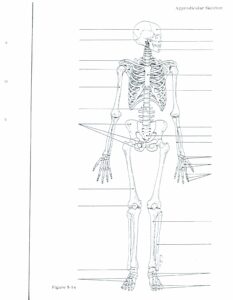
Part I
Click here for a diagram of the human skeleton. For this portion of the assignment, you must fill in the correct anatomical names for each of the major bones in the body. Briefly describe each identified bone on a document of 1–2 pages.
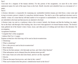
Part II
A forensic laboratory is responsible for examining any unidentified skeletal remains provided from a crime scene and trying to recreate a person’s identity by using those skeletal remains. The skull is one of the significant structures used to identify victims of a crime that has left them unable to be recognized or unidentifiable. It is essential to know that teeth are embedded in the head and that muscles are attached to the head.
A forensic team was sent to a warehouse where a fire occurred. Initially, the firemen said that the building was empty, but on the final walk-through of the building, they discovered what appeared to be burned human remains. The forensic team gathered all of the burned victim’s bones, tissue, and other pieces of clothing and took them to the laboratory for investigation.
Assignment Guidelines
• Complete Part I of the assignment.
• Address Part II in 3–4 pages:
Explain in detail how the following will be used in facial reconstruction:
Odontology
What is odontology?
How is this process used in facial reconstruction?
Bone formation
What are the three primary cells that make up bone, and what is their function?
What information can be obtained from the skeleton about growth?
How many bones are in the skull (face/head), and how are they important?
Muscles
What are the major facial powers, and what do they control?
What can be learned about the person’s identification based on the forces?