Benign Endometrial Hyperplasia Diagnosis
Cheryl,
Great post! I agree with the description of the benign endometrial hyperplasia diagnosis. As you have explained, the thickened uterine walls signify a cell overgrowth. This cell overgrowth occurs due to an imbalance of the two primary hormones, progesterone and estrogen. Various reasons lead to hormonal imbalance, including menopause, perimenopause, therapies for hormone replacement, irregular cycles, medications that imitate estrogen, infertility, obesity, and polycystic ovary syndrome. An age beyond 35 increases the risk for endometrial hyperplasia, as with the 40-year-old patient (Singh & Puckett, 2020).
The main hormones that are implicated in the diagnosis are estrogen and progesterone. Excess production of estrogen is responsible for the condition because of insufficient progesterone. As you have explained, atrophy involves a reduction in cell size, while hyperplasia consists of an increase in the size of cells. Hormonal stimulation can lead to hyperplasia, while the opposite can lead to atrophy. As you have described in the post, dysplasia and hyperplasia differ. In addition to the differences you have noted, dysplasia can be permanent, while hyperplasia is usually temporary. Medical interventions are necessary to revert the structural change of cells, which occurs as dysplasia (NCH, 2020). I want to commend the explanation that you have provided to defend your response regarding neoplasia and hyperplasia. The discussion’s clarity makes it easy to understand your point of view. Great discussion!
References
NCH. (2020). Left ventricular hypertrophy. Retrieved from https://www.nchmd.org/education/mayo-health-library/details/CON-20374296
Singh, G., & Puckett, Y. (2020). Endometrial Hyperplasia. StatPearls Publishing LLC. Retrieved from https://www.ncbi.nlm.nih.gov/books/NBK560693/
ORDER A PLAGIARISM-FREE PAPER HERE
We’ll write everything from scratch
Question
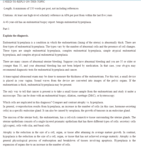
I NEED TO REPLY ON THIS TOPIC
-Length: A minimum of 150 words per post, not including references

Benign Endometrial Hyperplasia Diagnosis
Citations: At least one high-level scholarly reference in APA per post from within the last five years
A 40-year-old has an endometrial biopsy report: benign endometrial hyperplasia.
Part 1
Explain the diagnosis.
Endometrial hyperplasia is a condition in which the endometrium (lining of the uterus) is abnormally thick. There are four types of endometrial hyperplasia. The types vary by the number of abnormal cells and the presence of cell changes. These types are simple endometrial hyperplasia, complex endometrial hyperplasia, simple atypical endometrial hyperplasia, and complex atypical endometrial hyperplasia.
There are many causes of abnormal uterine bleeding. Suppose you have abnormal bleeding and you are 35 or older or younger than 35, and your abnormal bleeding has not been helped by medication. In that case, your ob-gyn may recommend diagnostic tests for endometrial hyperplasia and cancer.
A transvaginal ultrasound exam may be done to measure the thickness of the endometrium. For this test, a small device is placed in your vagina. Sound waves from the device are converted into images of the pelvic organs. If the endometrium is thick, endometrial hyperplasia may be present.
The only way to tell that cancer is present is to take a small tissue sample from the endometrium and study it under a microscope. This can be done with an endometrial biopsy, dilation, curettage (D&C), or hysteroscopy.
Which cells are implicated in this diagnosis? Compare and contrast atrophy vs. hyperplasia.
In general, overproduction results from hyperplasia, an increase in the number of cells (in this case, hormone-secreting cells) in a specific endocrine gland. It can also be caused by neoplasia, the growth of tumours in an endocrine gland.
The mucosa of the uterine body, the endometrium, has a cell-rich connective tissue surrounding the uterine glands. The uterine epithelium consists of a single-layered prismatic epithelium that has three different types of cells: secretory cells (glycogen), cells with cilia, and basal cells.
Atrophy is the reduction in the size of a cell, organ, or tissue after attaining its average mature growth. In contrast, hypoplasia is the reduction in the size of a cell, organ, or tissue that has not achieved average maturity. Atrophy is the general physiological process of reabsorption and breakdown of tissues involving apoptosis. Hyperplasia is the expansion of organs due to an increase in the number of cells.
How does dysplasia differ from hyperplasia?
Hyperplasia is an increase in the number of cells. It is the result of increased cell mitosis or division. The two types of physiologic hyperplasia are compensatory and hormonal. Compensatory hyperplasia permits tissue and organ regeneration. Hyperplasia is common in epithelial cells of the epidermis, intestine, liver hepatocytes, bone marrow cells, and fibroblasts. It occurs to a lesser extent in bone, cartilage, and smooth muscle cells. Hormonal hyperplasia occurs mainly in organs that depend on estrogen. For example, the estrogen-dependent uterine cells undergo hyperplasia and hypertrophy following pregnancy. Pathologic hyperplasia is an abnormal increase in cell division. A common pathologic hyperplasia in women occurs in the endometrium, called endometriosis.
Dysplasia generally refers to abnormal cellular shape, size, and organization changes. Dysplasia is not considered a faithful adaptation; it is thought to be related to hyperplasia and is sometimes called “atypical hyperplasia.” Tissues prone to dysplasia include cervical and respiratory epithelia. Dysplasia often occurs in the vicinity of cancerous cells, and it may be involved in the development of breast cancer.
Does hyperplasia lead to neoplasia? Defend your answer.
In some instances, pathological hyperplasia may progress to neoplasia. For example, hepatocellular adenoma or carcinoma is closely related to compensatory hyperplasia of hepatic parenchymal cells seen in cirrhotic livers of chronic alcoholics.
The progression begins with a mutation that makes the cell more likely to divide. The altered cell and its descendants grow and separate too often, a condition called hyperplasia. At some point, one of these cells experiences another mutation that further increases its tendency to separate; this cell’s descendants divide excessively and look abnormal, a condition called dysplasia. As time passes, one of the cells experiences yet another mutation, causing a very odd structure, loss of differentiation, and loss of contact between the cells; however, it is still confined to the epithelial layer from which it arose, which is called cancer in situ. In-situ cancer may remain contained indefinitely, but additional mutations may enable it to invade neighbouring tissues and shed cells into the blood or lymph; the tumour is considered invasive cancer (malignant). The escaped cells may establish new tumours (metastases) at other locations in the body.
References
Cellular adaptation – wikidoc. (2021). Retrieved 12 May 2021, from https://www.wikidoc.org/index.php/Cellular_adaptation
Endometrial Hyperplasia: Causes, Symptoms & Treatment. (2021). Retrieved 12 May 2021, from https://my.clevelandclinic.org/health/diseases/16569-atypical-endometrial-hyperplasia
Evolution of a Cancer. (2021). Retrieved 12 May 2021, from https://sphweb.bumc.bu.edu/otlt/MPH-Modules/PH/PH709_Cancer/PH709_Cancer5.html
(2021). Retrieved 12 May 2021, from https://quizlet.com/77740188/adaptations-hypertrophy-hyperplasia-atrophy-metaplasia-flash-cards/

