
Case Studies on Respiratory Disorders
Case 1: R.S.
Clinical Findings as a Consequence of COPD and Differences with Those of Emphysematous COPD
Clinical findings on the patient that are a consequence of COPD include PaCO2 of 60mmHg and PaO2. Increased PaCO2 seen in COPD is due to the inability of the lungs to fully exhale, causing retention of CO2 and consequent elevation of PaCO2. PaO2 is likely to fall in COPD due to lowered capacity of the lungs to transfer oxygen to the capillary space. Emphysema is a form of COPD that causes damage to the walls of the alveoli. Emphysematous COPD is characterized by profound elevation of PaCO2 due to airflow limitation and consequent CO2 trapping (Fazleen & Wilkinson, 2020).
Laboratory Results Interpretation
The acid-base disorder seen in the patient can be classified as respiratory acidosis. The patient’s ABG findings reveal a pH of 7.32 (Normal 7.35-7.45), indicating acidosis. The patient’s acid-base disorder can further be classified as respiratory acidosis due to the presence of PaCO2 elevation. The patient has a PaCO2 of 60mmHg (normal 38-42mmHg). The most likely cause of polycythemia is increased secretion of erythropoietin in response to chronic hypoxia.
The Rationale for Treating R.S. with Theophylline and a β2 Agonist
Theophylline and β2 agonists are effective in managing COPD. Theophylline use is merited in this case as it causes relaxation of the smooth muscles of the bronchioles with consequent bronchodilation. β-agonist also causes smooth muscle relaxation resulting in bronchodilation (Fazleen & Wilkinson, 2020). These two agents are used adjunctively for their synergistic effects on the bronchial smooth muscles.
Effects of the Respiratory Disease on the Patient’s Cardiovascular Functionalities
COPD has a negative impact on cardiovascular functionalities. COPD lowers the oxygen levels in the blood. The resulting hypoxemia may exacerbate left-side heart failure by placing additional stress on the heart. It can also result in pulmonary vasoconstriction with consequential right ventricular diastolic dysfunction (Fazleen & Wilkinson, 2020).
The Position in Which R.S. Will Have the Worst Ventilation-Perfusion Matching
Ventilation-perfusion matching will be worst when the patient is in the left lateral decubitus position. In this position, blood flows predominantly to the lower lung. There is also an increase in ventilation to the lower lung. This results in a significant reduction in ventilation-perfusion match in the non-dependent lung. In this case, the non-dependent lung is affected by pneumonia and COPD. This further worsens the ventilation-perfusion match on the non-dependent lung.
Case 2: T.V.
The Most Likely Cause of Distress in the Patient
The most likely cause of distress in the patient above is pulmonary embolism. Pulmonary embolism mostly occurs when a blood clot dislodges from the extremities, travels upwards, and embolizes in the pulmonary circulation. This causes disruptions in pulmonary circulation, with consequent manifestations of shortness of breath, sweating, and irregular heartbeat (Freund et al., 2022).
Diagnostic Findings That Would Help Confirm the Diagnosis
Several diagnostic workups are valuable in confirming pulmonary embolism (PE). Arterial blood gas analysis can reveal unexplained hypoxemia suggestive of PE. Elevations in brain natriuretic peptide and serum troponin levels are also suggestive of PE. Electrocardiographic abnormalities can also indicate the presence of PE. Chest radiography, computed tomography, pulmonary angiography, and pulmonary angiography can be used to visualize the respiratory system and detect the presence of an embolus (Freund et al., 2022). Other diagnostic techniques valuable in this case include magnetic resonance angiography, echocardiography, and compression ultrasonography.
Pathogenesis of the Hypoxemia in the Disorder
Hypoxemia in pulmonary embolism arises from disruption in the pulmonary circulation. Pulmonary embolism occurs when a clot breaks off from a primary site, mostly the lower extremities. This clot moves upward and is embolized in the pulmonary circulation. Larger emboli may obstruct the pulmonary arteries leading to pulmonary infarction. The resultant vascular obstruction impairs gas exchange leading to ventilation-perfusion mismatch and, consequently, hypoxemia.
Treating T.V.’s Respiratory Disorder
Management options for pulmonary embolism include anticoagulation therapy, supportive management, and reperfusion. Anticoagulation therapy using anticoagulants is the mainstay treatment option for pulmonary embolism. Low molecular weight heparin or fondaparinux are used in this regard. Oral anticoagulants and unfractionated heparin can also be used. Supportive measures include supplemental oxygen and volume resuscitation. Reperfusion strategies used include thrombolysis, surgical embolectomy, and catheter-directed treatment (Freund et al., 2022).
Case 3: R.J.
The Mechanism of Asthma and Airway Responses That Occur During Asthma Episodes
Asthma is a respiratory disorder characterized by airway inflammation. Asthmatic attacks often proceed the exposure to environmental triggers. Exposure to triggers such as animal dander and pollen triggers an inflammatory response in asthmatic patients due to bronchial hypersensitivity. This results in airway inflammation and increased mucous production with consequent narrowing and blockade of the airway (Papi et al., 2020). The manifestations of wheezing, cough, and shortness of breath result and are due to the narrowing of the airway and the body’s reflex to clear the airway. The three airway responses in an asthmatic episode are airway inflammation, smooth muscle contraction, and mucous hypersecretion.
Pharmacotherapeutic Options in Asthma Management
Several pharmacotherapeutic options are available in asthma management. Corticosteroids such as fluticasone, muscarinic antagonists such as ipratropium bromide, long-acting beta-agonists such as formoterol, leukotriene antagonists such as montelukast and methylxanthines such as theophylline are effective in managing asthma.
Significance of a PEFR at 60% Predicted
PEFR shows the amount of air that can be forcefully exhaled. PEFR at 60% predicted signifies that the patient’s airway is narrowing. It informs the need for corrective action. If spirometry is conducted at PEFR 60% predicted, the amount of air that the patient can forcefully exhale will only be 60% of the predicted amount.
Managing R.J.’s Problem and Monitoring Response to Therapy
The patient can be best managed by adding low-dose inhaled corticosteroids to the albuterol. Findings reveal poor response to the albuterol medications as demonstrated by continuous wheezing and an increase in asthmatic episodes prompting multiple inhaled albuterol use. Treatment response can be monitored by assessing symptoms recurrence, the need for inhaler use, night-time symptoms, and the activity level of the patients (Papi et al., 2020).
Preventive and Treatment Measures for Inclusion in R.J.’s Discharge Teaching
During discharge, the patient should be educated on conservative measures against the disease. This includes avoiding asthmatic triggers, environmental control to ensure that the environment is free from allergens, and education on bronchial thermoplasty, which is effective in preventing acute asthmatic attacks. The patient should also be educated on effective inhaler techniques to optimize the effect of the medications.
References
Fazleen, A., & Wilkinson, T. (2020). Early COPD: Current evidence for diagnosis and management. Therapeutic Advances in Respiratory Disease, 14, 175346662094212. https://doi.org/10.1177/1753466620942128
Freund, Y., Cohen-Aubart, F., & Bloom, B. (2022). Acute pulmonary embolism. JAMA, 328(13), 1336. https://doi.org/10.1001/jama.2022.16815
Papi, A., Blasi, F., Canonica, G. W., Morandi, L., Richeldi, L., & Rossi, A. (2020). Treatment strategies for asthma: Reshaping the concept of asthma management. Allergy, Asthma & Clinical Immunology, 16(1). https://doi.org/10.1186/s13223-020-00472-8
ORDER A PLAGIARISM-FREE PAPER HERE
We’ll write everything from scratch
Question 
Case 1
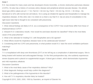
R.S. has smoked for many years and has developed chronic bronchitis, a chronic obstructive pulmonary disease (COPD). He also has a history of coronary artery disease and peripheral arterial vascular disease. His arterial blood gas (ABG) values are pH = 7.32, PaCO2 = 60 mm Hg, PaO2 = 50 mm Hg, HCO3- = 30 mEq/L. His hematocrit is 52% with normal red cell indices. He is using an inhaled B2 agonist and theophylline to manage his respiratory disease. At this clinic visit, it is noted on a chest x-ray that R.S. has an area of consolidation in his right lower lobe that is thought to be consistent with pneumonia.
Case Studies on Respiratory Disorders
Discussion Questions
1. What clinical findings are likely in R.S. as a consequence of his COPD? How would these differ from those of emphysematous COPD?
2. Interpret R.S.’s laboratory results. How would his acid-base disorder be classified? What is the most likely cause of his polycythemia?
3. What is the rationale for treating R.S. with theophylline and a B2 agonist?
4. What effects would his respiratory disease have on his cardiovascular function?
5. Considering both his COPD and pneumonia, in what position would R.S. have the worst ventilation-perfusion matching?
Case 2
T.V. is hospitalized with deep vein thrombosis (DVT) in her left leg as a complication of abdominal surgery. She is being treated with bedrest and anticoagulant therapy. On the third postoperative day, she suddenly experiences severe dyspnea and is placed on supplemental oxygen. A blood gas is drawn, which demonstrates hypoxemia and mild respiratory alkalosis.
Discussion Questions
1. What is the most likely cause of her respiratory distress? Why?
2. What diagnostic findings would help confirm this diagnosis?
3. What is the pathogenesis of the hypoxemia in this disorder?
4. How will T.V.’s respiratory disorder likely be treated?
Copyright © 2010, 2005, 2000, 1995 by Saunders, an imprint of Elsevier Inc.
Case 3
R.J. is a 15-year-old boy with a history of asthma diagnosed at age 8. His asthma episodes are triggered by exposure to cats and various plant pollens. He has been using his albuterol inhaler 10 to 12 times per day over the last 3 days and is continuing to wheeze. He normally needs his inhaler only occasionally (2 or 3 times per week). He takes no other medications and has no other known medical conditions. Physical examination reveals moderate respiratory distress with a respiratory rate of 32, oximetry 90%, peak expiratory flow rate (PEFR) 60% of predicted, and expiratory wheezing.
Discussion Questions
1. What is the underlying mechanism of R.J.’s asthma? What are the three airway responses that occur during an asthma episode?
2. In addition to bronchodilator therapy with intermittent albuterol, what other pharmacotherapy is important in the management of asthma?
3. What is the significance of a PEFR at 60% predicted? If spirometry were performed at this time, what would be the likely findings?
4. How should R.J.’s current problem be managed? What parameters should be assessed to monitor response to therapy?
5. In addition to reviewing appropriate drug therapy, what other preventive and treatment measures should be included in R.J.’s discharge teaching?
 Complete the order form and upload all of your files, including any instructions, rubrics, or other materials provided by your instructor.
 Complete the order payment after filling in the order form. We’ll receive your order and assign it to a writer.
 The assigned writer will complete your paper and forward it to our Editing Team for review and approval.
 Once approved by the editor, the completed paper will be uploaded to your account for you to download, review and approve.