Cell Biology
Be able to relate the structure, function, and location of the plasma membrane, cytoplasm, and nucleus. (3.1)
The plasma membrane is the outer boundary of the cell that constitutes a lipid bilayer composed of phospholipids and cholesterol. It covers the cell, separates the intracellular and extracellular substances, and controls materials’ entry and exit. Proteins attached to the lipid bilayer serve in intercellular communication, and the nucleus is placed at the center of the cell. A nuclear envelope, a double membrane with atomic pores, encloses it. The nucleus contains chromatin and nucleoli that consist of ribosomal RNA and proteins. The DNA in the middle is responsible for protein synthesis regulation and chemical reactions in the cell (Vanputte et al., 2020). The cytoplasm is a gel-like substance positioned between the plasma membrane and the nucleus. Key cellular functions such as protein folding and intracellular signaling are enabled by the physical and chemical properties of the cytoplasm (Luby-Phelps, 2013). Hire our assignment writing services in case your assignment is devastating you. We offer assignment help with high professionalism.
Be able to draw the fluid mosaic model of the plasma membrane, including membrane lipids (phospholipids, glycolipids) and membrane proteins (integral, peripheral, glycocalyx). Be able to explain the function of each. (3.3, 3.4)
(Vanputte et al., 2020). The figure above is a description of the fluid mosaic of the plasma membrane. The bilayer is made up of a dense liquid where molecules like proteins are suspended. It comprises membrane lipids and membrane proteins. Membrane lipids are phospholipids and glycolipids which form the lipid bilayer. Membrane proteins include the glycocalyx, integral, and peripheral proteins. The collection of glycolipids, glycoproteins, and carbohydrates on the outer surface of the plasma membrane forms glycocalyx. Essential proteins penetrate deep into the lipid bilayer, while peripheral proteins are located in the inner and outer surface of the bilayer.
The fluid mosaic model describes the plasma membrane structure as highly flexible in terms of its shape and composition. It allows the distribution of molecules within the plasma membrane, assembles phospholipids around damaged sites for repair, and allows the fusion of membranes with one another.
Be able to describe the structure of phospholipids and why they form a lipid bilayer, especially in relation to the terms hydrophobic and hydrophilic. (3.4)
The double membrane is due to the polar heads and nonpolar tails nature of phospholipid molecules. The charges are hydrophilic as they are attracted to water molecules and are exposed to the aqueous fluids in the extracellular and intracellular environments of the cell. The hydrophilic tails are not attracted to water molecules; they face each other in the interior of the plasma membrane.
List several functions of integral membrane proteins. (3.5)
Essential membrane proteins function as:
- Attachment proteins that allow attachment of cells to other cells or extracellular molecules.
- Marker molecules which help cells to recognize other cells or molecules.
- Transport proteins that enable the movement of molecules across the plasma membrane.
- Receptor proteins allow for the attachment of specific substances to the outer surface of the cell.
- Enzymes catalyze chemical reactions in the inner and outer surfaces of the plasma membrane.
Compare and contrast three types of cell junctions (tight junctions, desmosomes, gap junctions). Be able to list locations where each is found. (on your own) (Figure 4.2, pg 110-111)
Types of cell junctions include tight junctions, desmosomes, and gap junctions. Tight junctions are intercellular between epithelial and endothelial cells, creating a semipermeable diffusion fence that prevents the mixture of apical and basolateral components. They are also essential in cell growth regulation and differentiation (Balda and Matter, 1998). Desmosomes are intercellular junctions that function as adhesive complexes and cell-surface attachment sites for intermediate filaments to the plasma membrane. They maintain cell integrity and regulate cell behavior through the transduction of intracellular signals (Kowalczyk et al., 1998). Finally, gap junctions are intercellular channels that occur across adjacent cells and allow for direct cell-to-cell transfer of ions and small molecules (Goodenough and Paul, 2009).
Define selectively permeable. What kinds of molecules can cross the membrane without help? (3.6)
Selective permeability is the ability of the plasma membrane to allow the passage of certain molecules only through it. The intracellular and extracellular environment has different compositions of molecules that the cell’s survival is dependent on. For instance, potassium ions are highly concentrated intracellularly, while sodium ions are highly concentrated extracellularly.
Define the term concentration gradient. What is needed to move molecules against their concentration gradient? (3.6)
The concentration gradient refers to the concentration difference between two points over the distance between these two points. For movement to occur against the concentration gradient, solutes need to be unequally distributed in a solvent. The solutes will diffuse from where they are highly concentrated to where they are lowly focused.
Distinguish between the different transport processes: simple diffusion, facilitated diffusion, osmosis, and active transport (primary and secondary). Include the direction of the vehicle in relation to the concentration gradient, use of protein carrier or pump, use of ATP, and examples of each process, including what kinds of molecules are moved by each. (making a table will help) (3.6)
Simple diffusion is the movement of molecules along a concentration gradient. Molecules transported include soluble lipid molecules, and an example is the diffusion of steroid hormone into the lipid bilayer.
Facilitated diffusion involves the movement of substances through pores of the membrane with the help of carrier proteins. ATP is not used. Examples include the movement of glucose into muscle cells.
Osmosis is the movement of water through a selectively permeable membrane from low to high concentrations, for instance, the movement of water from the intestines into the blood.
Active transport is the movement of substances to a higher concentration across the membrane through ATP-powered pumps, as in the case of the active transportation of calcium ions.
Secondary active transport is the movement of ions (such as sodium ions) through active transport against a concentration gradient and with ATP, for example, the movement of glucose from the intestinal lumen into epithelial cells.
Describe the workings of the sodium potassium pump. What is its goal? List the steps involved in its action, including what ions are pumped and where. (3.6)
The work of the sodium-potassium pump is to move sodium ions (Na+) out of the cell and potassium ions (K+) in. Firstly, three Na+ and adenosine triphosphate (ATP) bind to the sodium-potassium pump. ATP is broken down into adenosine diphosphate and phosphate to release energy, which changes the shape of the pump. Na+ then moves into the extracellular fluid across the membrane, and then two K+ bind to the pump, releasing the phosphate molecule. The pump changes shape to transport K+ into cytoplasm across the membrane and allows for the binding of Na+ and ATP.
Define tonicity and three states of tonicity (isotonic, hypertonic, and hypotonic). Be able to predict what will happen to a red blood cell when placed in each of the three solutions. (3.6)
Tonicity refers to the constant shape of a cell whereby its internal tension is maintained. States of tonicity include isotonic, hypertonic, and hypotonic. Isotonic solutions have equal solute concentration and do not cause shrinking or swelling of cells such as red blood cells. Hypertonic solutions have greater solute concentrations and high osmotic pressure that cause the shrinking of cells. In contrast, hypotonic solutions are dilute with lower osmotic pressure and cause swelling of a cell.
Predict the osmotic direction based on solute and water concentrations. (3.6)
Osmotic pressure is responsible for the osmotic direction based on solute and water concentration. For instance, water molecules move from a cell placed in a hypertonic solution into a lowly concentrated solution, resulting in creation.
Define endocytosis and exocytosis. Compare and contrast phagocytosis with pinocytosis. Fig 3.12, 3.13, and 3.14 are helpful. (on your own) (3.6)
Endocytosis is the transportation of substances through a cyst formed by the plasma membrane and requires ATP. Phagocytosis involves taking in cells and solid particles, while pinocytosis constitutes taking in molecules dissolved in liquids. Exocytosis, however, consists of the fusion of the particles into secretory vesicles that release these particles out of the cell. It also requires ATP.
For each of the following organelles, be able to list its function and recognize it in a diagram of the cell: cytoplasm, cytosol, cytoskeleton, extracellular matrix, Golgi apparatus, mitochondria, lysosome, nucleoli, nucleus, peroxisome, ribosome, rough endoplasmic reticulum, smooth endoplasmic reticulum. (on your own) (3.7, 3.8)
Cytoplasm functions to enable key cellular functions such as protein folding and intracellular signaling through its physical-chemical properties.
The cytosol is the fluid part of the cytoplasm that dissolves ions and proteins, while the cytoskeleton supports the cell, holds the organelles, and controls their shape and movement.
The extracellular matrix contains protein fibers, fluids, and other molecules that enable the skin to bear damages like punctures and bones to withstand the weight.
Golgi apparatus is the center for packaging and distributing proteins and lipids manufactured in the rough endoplasmic reticulum.
Lysosomes harbor digestive enzymes.
Peroxisomes are the center for the degradation of lipids and amino acids and hydrogen peroxide breakdown.
Mitochondria is the site for major ATP synthesis in the presence of oxygen.
The nucleus controls the cell and regulates the synthesis of proteins, while the nucleolus contains ribosomal RNA and proteins.
Ribosomes form sites for protein synthesis.
The rough endoplasmic reticulum performs protein synthesis, while the smooth endoplasmic reticulum is a manufacturer of lipids and carbohydrates, a detoxifier of harmful chemicals, and calcium storage.
Compare and contrast the general function of microvilli, cilia, and flagella (on your own) (3.8)
Microvilli increase the surface area for absorption and secretion of the plasma membrane. They are also modified to sensory receptors—Cilia transport substances over the surface, while flagella move spermatozoa in humans.
Be able to list the phases of the cell lifecycle in order, as well as what major events occur in each. Fig 3.31 (on your own) (3.10)
Stages of the cell cycle include interphase, prophase, metaphase, anaphase, telophase, and cytokinesis. During interphase, digestive enzymes are secreted, DNA is replicated, and the centrioles are duplicated. In prophase, the chromatin condenses, chromosomes replicate, centrioles move to opposite ends of the cell, spindle fibers begin to form, and the nuclear envelope begins to disappear. The chromosomes then align towards the center in metaphase. Separation of chromatids follows at the onset of anaphase. The identical sets of chromosomes move to the opposite poles, and the division of the cytoplasm begins. In telophase, a nuclear envelope is formed around the two separate chromosomes while they uncoil to resemble the genetic composition of the parent cell. Lastly, during cytokinesis, the cytoplasm is divided to produce two identical daughter cells.
Compare and contrast DNA replication, transcription, and translation, including the function of each, where it takes place in the cell, and which organelles are involved. Be able to trace the pathway flow of information between these steps. (3.9)
DNA replication is the separation of the two strands of a DNA molecule, whereby one serves as a template for producing new complementary nucleotide strands. Transcription involves the synthesis of mRNA, tRNA, and rRNA. DNA strands unwind through nucleotide base pairing to produce pre-mRNA. Also, introns are removed while exons become spliced, and the ends of mRNA are modified during the post-transcriptional process. The translation is the synthesis of proteins depending on the sequence of codons of mRNA, which requires tRNA and rRNA; the anticodons of tRNA and mRNA bind, causing amino acids to join, thus forming a protein. Additionally, during post-translation, the proproteins (proenzymes and enzymes) become proteins.
References
Balda, M. and Matter, K. (1998). Tight junctions. Journal of Cell Science, 111(5), pp.541-547.
Goodenough, D. and Paul, D. (2009). Gap Junctions. Cold Spring Harbor Perspectives in Biology, [online] 1(1), pp.a002576-a002576. <http://sci-hub.se/10.1101/cshperspect.a002576>
Kowalczyk, A., Bornslaeger, E., Norvell, S., Palka, H. and Green, K. (1998). Desmosomes: Intercellular Adhesive Junctions Specialized for Attachment of Intermediate Filaments. International Review of Cytology, [online] pp.237-302. <http://sci-hub.se/10.1016/S0074-7696(08)60153-9>
Luby-Phelps, K. (2013). The physical chemistry of cytoplasm and its influence on cell function: an update. Molecular Biology of the Cell, [online] 24(17), pp.2593-2596. <http://sci-hub.se/10.1091/mbc.e12-08-0617>
Vanputte, C., Regan, J., Russo, A., Seeley, R., Stephens, T. and Tate, P. (2020). Seeley’s Anatomy & Physiology. 12th ed. New York: McGraw-Hill Education.
ORDER A PLAGIARISM-FREE PAPER HERE
We’ll write everything from scratch
Question
Membrane Transport
This activity is designed to be completed BEFORE the Ch3: Cell biology PowerPoint and BEFORE the Ch3: Cell Biology lecture videos. All of the information you need to answer the questions is present in the models (the pictures for each section). You should not need to reference texts, notes, or the internet. There are many correct answers to some questions.

Cell Biology
Model 1: Active Versus Passive Transport
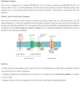
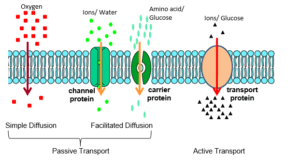
Body fluids are solutions of water and dissolve solutes (ions, glucose, amino acids, etc.) that surround our cells. The plasma membrane is a selectively permeable barrier that allows the passage of some ions and molecules but prevents the course of many molecules essential for life. The selection of these molecules through the semipermeable plasma membrane is necessary for life. Active transport and passive transport are two transport mechanisms responsible for moving substances across the plasma membrane.
Questions
- On the model: for each transport process, label which side of the membrane has a high solute concentration (amount) and which side has a low solute concentration.
- Transport mechanisms are determined by how solutes move in regard to their concentration gradient (i.e., high to low, low to high).
- Examine the model. In passive transport, how do solutes move along their concentration gradient?
- In active transport, how do solutes move along their concentration gradient?
- The movement of molecules is often stated as moving ‘up’ or ‘down’ the concentration gradient. Fill in the blanks:
Passive transport moves solutes _____________ its concentration gradient, and active vehicle moves solutes ______________ its concentration gradient.
- Cells use energy in the form of ATP to do work. Which transport process(es) will use ATP? Write a complete sentence to explain why.
- Diffusion is the random movement of molecules until equilibrium is reached.
- Examine model one. What type of transport (active or passive) is diffusion? Explain why.
- Examine the model. List the two types of diffusion.
- Which process(es) involves transport directly through the phospholipid membrane?
Which process(es) requires a protein channel/carrier for transport?
- Solutes being transported by simple diffusion are impermeable/permeable (circle one) to the plasma membrane. Solutes being transported by facilitated diffusion are impermeable/permeable (circle one) to the plasma membrane.
- The several factors influence rate of diffusion through a membrane. Managers have each membrane of the team take turns to predict which condition will allow distribution to occur faster:
- Size of molecules – smaller or larger?
- Temperature of molecules – cooler or warmer?
- Membrane surface area – less or more?
- Membrane permeability – less or more?
- The steepness of concentration gradient – less different or more different?
- The Na-K pump actively transports potassium into the cell and sodium out of the cell across the plasma membrane. Draw a cell showing the Na and K distribution (high/low) for both the inside and outside of the cell.
- A cell uses valuable energy to run active transport. Why does a cell need active transport? Make sure the explanation includes two examples of when active transport is used in the body.
Model 2 – Osmosis
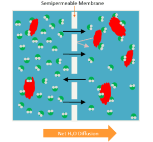
Osmosis is the diffusion of water through a semipermeable membrane to maintain the equilibrium of solutes on both sides of the membrane. It is a solute imbalance that causes water to move across the plasma membrane. The saying goes: “Water follows salt (solute).”
Questions for Model 2
- In model 2: Label the water and solute molecules.
- Based on the model:
- a) How does water move in regard to its concentration
- b) How does water move in regards to the solute concentration
- What causes osmosis, an imbalance in water concentration or solute concentration (Tricky question!)?
- Water, a polar molecule, needs to constantly move in and out of the cell at rapid speed to maintain solute equilibrium. Decide the specific type of transport used for osmosis. Explain your answer using the terminology learned in model one.
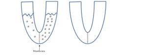
- The solutions in the two arms of this U-tube are separated by a membrane that is impermeable to glucose. The left arm contains six glucose molecules, and the right arm contains 14 glucose molecules. Predict the solution volume and number of glucose molecules in each component at the end of equilibrium.
- At equilibrium, how does the glucose concentration on the left side compare to the right side? [Remember concentration is the amount of solute per water].
- In another scenario. The solutions in the two arms of this U-tube are separated by a membrane that is permeable to sucrose. The left arm contains two sucrose molecules, and the right arm contains 12 sucrose molecules. Predict the solution volume and sucrose molecules in each component at the end of equilibrium.
Model 3 – Tonicity
- Based on model 3, how does the impermeable solute concentration in the solution (fluid surrounding the cell) compare to the inside of the cell?
Hypotonic =
Isotonic =
Hypertonic =
- Based on your knowledge of osmosis (refer to model 2), predict the net water movement for each tonicity solution.
Hypotonic =
Isotonic =
Hypertonic =
- Tonicity is the ability of a solution to change the shape/tone of a cell by altering its internal water volume. Based on your answer above, predict how RBC shapes after it has reached equilibrium with each solution. Manager: have team members take turns predicting the final shape. Then, as a group, draw a representative final RBC.
- Use model 3 and question 13 &15 responses to answer this question. Is tonicity due to the solution containing a permeable or impermeable solute? Justify your answer.
- Normal plasma (blood fluid) osmolarity is about 300 mm/kg. An older woman with diabetes has elevated blood glucose levels that raise her blood osmolarity to 310 m/kg. In this diabetic patient, predict the water movement, if any, of the extracellular fluids and the shape of her red blood cells. [Hint: the diabetic cells are initially in an isotonic fluid that is the same as normal blood.]
- A patient was in a serious accident that caused a lot of blood loss. In an attempt to replenish body fluids, distilled water (distilled water means NO solutes) with a pH of 7.0, equal to the volume of blood lost, is transferred directly into the patient’s veins. What will be the most probable result of the person’s red blood cells from this transfusion? Explain your answer using membrane transport terminology learned in these models.
- Recap: fill-in the below summary table.
| Transport Mechanism | Gradient Direction | Protein Carrier? | ATP needed? | Molecule Examples |
| Simple Diffusion | ||||
| Facilitated Diffusion | ||||
| Osmosis | ||||
| Active Transport |


