
The Cardiovascular System
1. a). Auscultation of the valves
The tricuspid valve is auscultated in the fourth intercostal space on the sternal left upper margin. The bicuspid (mitral) valve is heard on the fifth intercostal space on the left, on the medial side of the midclavicular line. The aortic valve is auscultated on the intercostal second space on the sternal upper right margin. The pulmonary valve is auscultated on the sternal upper left margin intercostal double space. (Amerman, 2021).
Are you looking to obtain a unique copy of “The Cardiovascular System” ? Contact us.
b). Newborn auscultation of abnormal heart sound
The ideal spot for auscultating abnormal heartbeat is from the left lower sternal margin to the right lower sternal margin. The reason is that the interventricular septum, which separates the atrioventricular valves, overlies the ventricular septal defect (VSD). (Amerman,2021).
Normal heart sounds and their causes.
The average heart sounds are S1 and S2. The first heart sound, or “lub,” is called S1 because the atrioventricular valves, bicuspid and tricuspid, close. The second heart sound, or “dup,” is called S2. Because of the closing semilunar valves, that is,
of the aortic and pulmonary valves.
3. a). Ventricular septal defect
Following the ventricular septal defect, I anticipate the blood will travel from the left ventricle to the right ventricle during systole. This is because the left ventricle has a higher blood pressure than the right ventricle.
b). Blood pressure and heart resistance
Blood flows down its gradient of resistance (pressure) or into an open area in which there is the minimum amount of pressure. The blood pressure in the left ventricle is higher than in the right ventricle. Such a situation occurs due to the afterload of the right ventricle being less than its afterload (Pappano & Wier, 2018). The arteries of pulmonary circulation produce lower resistance than the blood vessels of the systemic circulation, which accounts for the afterloads variation.
Some anatomical heart abnormalities cause the mixing of deoxygenated and oxygenated blood. They include Atrial septal defect (ASD) and Patent ductus arteriosus (PDA). Usually, very shortly after delivery, the ductus arteriosus, which joins the aorta with the fetus’s pulmonary trunk, shuts. If it doesn’t close, though, it stays patent or open. Because of a PDA, some of the aorta’s oxygenated blood can go back to the pulmonary trunk and combine with deoxygenated blood, which is then transported to the lungs. Thence, the amount of blood that travels to the lungs rises. (Pappano & Wier, 2018). ASD refers to a wall defect that occurs between the upper two chambers. The inferior and superior cava typically transport systemic deoxygenated blood to the right atrium, while the left atrium receives oxygenated blood from the pulmonary veins. Some oxygenated blood returns to the heart’s right side when there is a gap within the atria. Although it already contains a lot of oxygen, it returns to the lungs (Pappano & Wier, 2018). As a result, the amount of blood that travels to the lungs rises significantly.
Blood cell path
Deoxygenated blood from the inferior bodily parts is emptied into the inferior vena cava. The right atrium receives the inferior vena cava’s drainage. The tricuspid valve controls right ventricular blood flow. It travels through the tricuspid valve and enters the pulmonary artery before getting to the lungs. Within the lungs, the blood separates into the numerous pulmonary capillaries, blowing out carbon dioxide and taking in oxygen. The pulmonary vein carries blood into the left atrium, where it is fully oxidized before being pumped into the left ventricle via the bicuspid valve. The aortic valve, which transports blood rich in oxygen to all bodily areas, is opened by the left ventricle’s muscular contractions, which force oxygenated blood through it. (Michael & McFarland, 2020). Deoxygenated blood is then pumped back to the IVC.
The superior vena cava allows deoxygenated blood from the upper body parts to reach the right atrium. It travels through the right auricle to the right ventricle through the suitable atrioventricular valve. The right ventricle’s contraction forces the pulmonary semilunar valve to open. Blood can flow into the pulmonary trunk from the tricuspid valve. Blood travels through the left and right pulmonary arteries to the lungs, absorbing oxygen and exhaling carbon dioxide. Blood leaves the lungs and travels through the pulmonary veins to the left auricle. Left ventricular tension forces the semilunar valve of the aorta to open. For blood to enter the ascending aorta, the aortic valve must open. The aorta delivers blood to all internal organs, unloads oxygen, and takes in carbon dioxide (Michael & McFarland, 2020). This carbon-rich blood is then pumped back to the superior vena cava.
Oxygenated and deoxygenated blood
The left ventricle contains oxygenated blood. Since oxygen-rich blood is transported to the left atrium from the lungs, the left ventricle subsequently circulates blood from the left atrium throughout the body, supplying every organ with oxygen-rich blood (Herring & Paterson, 2018). On the other hand, the right ventricle contains deoxygenated blood. Because the right atrium takes up low-oxygen blood from the body, this is vein-derived blood (Herring & Paterson, 2018). The right ventricle subsequently pumps the blood from the right atrium into the lungs, where carbon dioxide and oxygen are exchanged.
Systemic cardiac output
Systemic cardiac output refers to the process by which blood flows in the body, which is determined by the amount of blood pumped by the heart every minute, hence supplying the nervous system and other crucial parts with blood. When exercising, the body’s need for oxygen changes and modifies both stroke volume (SV) and heart rate (HR), affecting the systemic cardiac output (Amerman, 2021). As a result, a complicated process encompassing the autonomic nervous system, paracrine signal transduction, and endocrine system controls the systemic cardiac output.
Venous return, pulse rate, and contraction force are three elements that affect cardiac output. Venous return refers to the blood volume that passes through the veins and into the heart in a minute. The cardiac output and venous return are equal after a specific time; cardiac output rises with an increased heart rate (Amerman, 2021). As the force of contraction grows, so does the cardiac output and stroke volume.
References
Amerman, E. C. (2021). Human anatomy & physiology. EBONY HOLT.
Herring, N., & Paterson, D. J. (2018). Levick’s introduction to cardiovascular physiology. CRC Press.
Michael, J., & McFarland, J. (2020). Another look at the core concepts of physiology: revisions and resources. Advances in Physiology Education, 44(4), 752-762. https://doi.org/10.1152/advan.00114.2020
Pappano, A. J., & Wier, W. G. (2018). Cardiovascular physiology-e-book. Elsevier Health Sciences.
ORDER A PLAGIARISM-FREE PAPER HERE
We’ll write everything from scratch
Question 
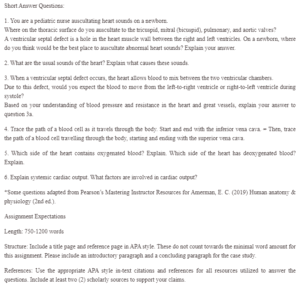
Short Answer Questions:
1. You are a pediatric nurse auscultating heart sounds on a newborn.
Where on the thoracic surface do you auscultate to the tricuspid, mitral (bicuspid), pulmonary, and aortic valves?
A ventricular septal defect is a hole in the heart muscle wall between the right and left ventricles. On a newborn, where do you think would be the best place to auscultate abnormal heart sounds? Explain your answer.
The Cardiovascular System
2. What are the usual sounds of the heart? Explain what causes these sounds.
3. When a ventricular septal defect occurs, the heart allows blood to mix between the two ventricular chambers.
Due to this defect, would you expect the blood to move from the left-to-right ventricle or right-to-left ventricle during systole?
Based on your understanding of blood pressure and resistance in the heart and great vessels, explain your answer to question 3a.
4. Trace the path of a blood cell as it travels through the body. Start and end with the inferior vena cava. = Then, trace the path of a blood cell travelling through the body, starting and ending with the superior vena cava.
5. Which side of the heart contains oxygenated blood? Explain. Which side of the heart has deoxygenated blood? Explain.
6. Explain systemic cardiac output. What factors are involved in cardiac output?
*Some questions adapted from Pearson’s Mastering Instructor Resources for Amerman, E. C. (2019) Human anatomy & physiology (2nd ed.).
Assignment Expectations
Length: 750-1200 words
Structure: Include a title page and reference page in APA style. These do not count towards the minimal word amount for this assignment. Please include an introductory paragraph and a concluding paragraph for the case study.
References: Use the appropriate APA style in-text citations and references for all resources utilized to answer the questions. Include at least two (2) scholarly sources to support your claims.