
Documentation of an Assessment of the Head, Ears, and Eyes
Subjective Data
The patient is Mrs.L.K., a 35-year-old black female presenting with a history of head injury for eight hours. She was showering when she toppled off, fell, and hurt the back of her head. She then experienced pain that was sharp, constant, and radiating to the face and the neck. It was accompanied by mild bleeding and swelling from the occipital region, nausea, and hotness. It was also associated with loss of consciousness, hearing loss, vision loss, blurry vision, otalgia, and eye pain. There was no bleeding or discharge from the ears, eyes, nose, or mouth. The patient reports no relieving factors, and she used paracetamol to relieve the pain, but it remained constant. The patient grades the severity of the pain at 9/10. Do you need help with your assignment ? Contact us at eminencepapers.com.
Objective Data
Upon inspection, the head was mildly bleeding from the occipital region; the hair was equally distributed on the scalp. There were no contusions, lacerations, or scars. On palpation, there was tenderness in the occipital and parietal regions and swelling of approximately 2 cm by 2 cm in the occipital region. On eye examination, the eyes were symmetrically equal; there were no scars, crusting, swelling, redness, ptosis, discharge, or bleeding. The eyelashes and eyebrows were normal. The visual acuity was 6/6; the extraocular movements were intact. The conjunctiva was not injected, the lens and the cornea were clear, and the pupils were round and reactive to light. On-ear examination, the tragus, and the pinna were normal; there were no impacted wax, foreign body, redness, discharge, swelling, bleeding, or scars, and the external auditory meatus was clear and grey. The hearing tests were all normal.
Actual or Potential Risks
There is a potential risk of increased intracranial pressure due to increased CSF or brain hemorrhage. Visual alterations brought on by elevated intracranial pressure may be linked to a history of head injury (Sullivan, 2018). The patient might develop infections such as meningitis or encephalitis. Moreover, the patient is at a higher risk of convulsions due to the elevation of CSF and infections (Naoya, 2021). Lastly, the patient is at risk of persistent headaches due to tension and injury alterations of the brain.
References
Naoya, T. (2021). Complications Arise During Traumatic Brain Injury—Journal of Neurology, 12(11), 565.
Sullivan, D. D. (2018). Guide to clinical documentation. FA Davis.
ORDER A PLAGIARISM-FREE PAPER HERE
We’ll write everything from scratch
Question 
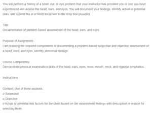
You will perform a history of a head, ear, or eye problem that your instructor has provided you or one you have experienced and assess the head, ears, and eyes. You will document your findings, identify actual or potential risks, and submit this in a Word document to the drop box provided.
Documentation of an Assessment of the Head, Ears, and Eyes
Title:
Documentation of problem-based assessment of the head, ears, and eyes.
Purpose of Assignment:
I am learning the required components of documenting a problem-based subjective and objective assessment of a head, ears, and eyes. Identify abnormal findings.
Course Competency:
Demonstrate physical examination skills of the head, ears, eyes, nose, mouth, neck, and regional lymphatics.
Instructions:
Content: Use of three sections:
o Subjective
o Objective
o Actual or potential risk factors for the client based on the assessment findings with description or reason for selecting them.