
Asthma Management
The case presented is of a 42-year-old patient called J.S., who was brought to the emergency room with complaints of wheezing. The wheezing was unresponsive to the salmeterol/ fluticasone combination he had been using. The patient was also unable to lie down and had difficulty breathing, as evidenced by the use of accessory muscles. He was also anxious. A preliminary laboratory investigation reveals a B.P. of 152/84, a heart rate of 124 BPM, and a temperature of 110.04F. His ABG findings revealed a pH of 7. 31. PaCO2 of 48, PaO2 of 55, and HCO3 of 26.
Do you need an original copy of “Asthma Management” ? Contact us.
The symptoms that the patient presented with are consistent with the symptoms of asthma. Asthma is a chronic respiratory disease characterized by inflammation of the airway. Airway narrowing and blockade become apparent as the disease becomes more severe. The critical feature of asthma is airway hyperresponsiveness and subsequent inflammation that leads to the manifestation of symptoms. Asthma remains a common respiratory pathology affecting up to 20% of people in developed countries. Asthma has been implicated in significant morbidity and mortality.
Causes of Asthma
Asthma is a multifactorial respiratory pathology linked to several causal factors. Genetic predisposition to the disease, personal or family history of atopy, and exposure to triggers have been linked to asthma development. The risk for asthma is up to threefold higher in persons with close relatives with the disease. Asthma is also more common in atopic individuals or those with a history of atopy. Asthma is likely to develop in individuals with a propensity to be allergic to common environmental allergens such as hay and animal dander. Asthma has also been linked with active or secondhand exposure to tobacco smoke, particulate matter, and inflammatory gases.
Exposure to environmental triggers is a leading cause of asthmatic attacks among susceptible individuals. These triggers include viral respiratory tract infection, insect or chemical fumes, environmental allergens such as hay, pollen, animal dander, cold air, tobacco smoke, and certain drugs such as aspirin and beta blockers. Other trigger factors include obesity, exercise, gastroesophageal reflux disease, stress, other emotional factors, and chronic sinusitis.
Symptomatic presentations seen in asthma often follow a trigger. Due to hyperresponsiveness of the airway, exposure to a trigger factor usually elicits an inflammatory response, with mucus production being apparent. An asthmatic attack usually involves inflammatory infiltrates of eosinophils, mast cells, neutrophils, and monocytes. The subsequent obstruction seen as the disease progresses results from bronchoconstriction resulting from a vascular response to the inflammatory process, mucus plug formation resulting from mucus hypersecretion, and inflammatory cell infiltration. These are the disease’s hallmarks and contribute to symptom manifestation and chronic disease (Bush, 2019). These symptoms are reversible with treatment. Over time, irreversible changes that highlight poorly controlled diseases may become apparent. These changes include collagen deposition, basement membrane thickening, epithelial desquamation, and airway remodelling due to smooth muscle hyperplasia and hypertrophy. The prognosis is poor when irreversible changes kick in.
Symptoms
Patients with asthma present with symptoms like shortness of breath, wheezing, and cough; additionally, chest tightness che, st pain, and insomnia due to trouble sleeping are also common complaints in primary care. Chest tightness in the disease indicates an acute exacerbation of the disease. Diurnal variations in symptom presentation occur with the disease and may be reported by the patients (He et al., 2019). Asthma symptoms tend to worsen at night, often causing sleeping disturbances.
Diagnosis of asthma follows a thorough evaluation and history-taking to ascertain the presence of risk factors, existing symptoms of the disease, and laboratory findings. History taking informs on the presence or absence of predisposing factors such as a history of atopy in the patient and or their close relatives, genetic predisposition to the disease, and history of an asthmatic attack in the patient. Laboratory findings give information on urea and electrolyte levels in the blood, ABGs, eosinophilia, and ECG readings. Urea and electrolytes are markers of kidney function. They are valuable for patients receiving high doses of terbutaline or on repeat doses since terbutaline causes a potassium shift and may result in transient hypokalemia. ABG reveals the presence of respiratory acidosis and hypoxemia and may guide the treatment approach to be used on the patient. Electrocardiograph readings may show the presence of sinus tachycardia that may sometimes accompany the disease and may also be caused by antiasthmatic medications such as theophylline and albuterol. In exceptional circumstances where the patient is unresponsive to therapy and has recurrent symptoms, a chest C.T. scan may be warranted to determine the presence of an underlying pathology that may be causing the symptoms.
Status asthmaticus defines asthma that is unresponsive to conventional approaches to therapy. This condition is a medical emergency characterized by hypercarbia, hypoxemia, and secondary respiratory failure. The sudden onset asthma exacerbation subset of status asthmaticus presents with severe deterioration and symptom flares over a few hours. This condition results from sudden and massive exposure to environmental triggers that the patient is otherwise allergic to. These triggers include but are not limited to animal dander, pollen, and hay.
The patient presented was brought to the hospital with wheezing that was unresponsive to the salmeterol/ fluticasone combination that the patient had been taking for his asthma. Additionally, the patient had dyspnea. His vital readings revealed a high pulse rate of 124bpm (normal range:60-100), a high B.P. of 152/84 (average range:120-130/80-84), and a respiratory rate of 42 breaths per minute (normal range: 12-16). These findings show that the patient was hyperventilating. Hyperventilation is a reflex response to hypercarbia—hyperventilation attempts to remove the excess CO2 accumulated in the systemic circulation.
ABG findings on the patient revealed that the patient had acidosis, as evidenced by a low blood pH of 7.31 (normal range: 7.35-7.45). The patient also had a PaCO2 of 48 (normal range: 38-45mmHg). This reaffirms that the patient had hypercarbia. His PaO2 was 55 (normal range: 75-100). This indicates that the patient was hypoxemic, as he had abnormally low oxygen levels in his bloodstream. The presence of hyperventilation, as evidenced by the patient’s respiratory rates and unusually high levels of CO2 as evidenced by PaCO2, may indicate a secondary respiratory failure. The presence of hypoxemia, hypercarbia, and secondary respiratory failure in an asthmatic who is unresponsive to conventional therapies are reflective of status asthmaticus (Agnihotri & Saltoun, 2019). The patient is, therefore, suffering from this condition.
The sudden onset asthma exacerbation seen in the patient J.S. may have been caused by massive exposure to dust or pollen. As indicated in the case study, the patient has mild asthma and is allergic to pollen and dust. Huge exposure to these allergens may have triggered an inflammatory cascade that resulted in acute exacerbations of his disease. Massive exposure to allergens and preexisting asthma disease are the two major causal factors for sudden onset asthma exacerbation subset of status asthmaticus.
Treatment
The first approach to treating the patient presented in the case study was to stabilize him using oxygen and intravenous fluids. J.S. was started on 4 L oxygen by nasal cannula and intravenous (IV) D5W at 75 mL/hr at the emergency department. Oxygen was used in this case to offset the hypoxemia that was seen in the patient. D5W, on the other hand, was used to offset the respiratory acidosis that was seen in the patient (Le Conte et al., 2019). these early interventions may stabilize the patient and prevent further deterioration.
Pharmacological interventions remain the mainstay modalities of treating status asthmaticus. Beta-agonists, anticholinergics, and corticosteroids are the primary pharmacological approaches used in this disease. Short-acting inhaled beta-agonists are the first line in this regard. Albuterol is the preferred agent due to its high selectivity and longer duration of pharmacologic action. Prednisone is the corticosteroid of choice in this condition, often given in doses of between 150mg and 225mg over 24 hours. Intravenous methylprednisolone at a dose of 60-125mg every 6 hours can also be used in the emergency treatment of the disease. Anticholinergic medications such as ipratropium bromide can offer significant benefits in patients experiencing bronchospasms induced by the primary disease. Magnesium sulfate is also effective in this regard, as it inhibits calcium-induced bronchoconstriction. Intravenous magnesium sulfate has been used adjunctively with beta-agonists (Papi et al., 2020). J.S. can benefit considerably from these pharmacological approaches.
Finally, J. S. is a known asthmatic who presented with symptoms that are similar to symptoms presented in status asthmaticus. He presented with hypoxemia, hypercarbia, and secondary respiratory failure, which are all characteristic of sudden onset asthma exacerbation, a subset of status asthmaticus. Just as seen in this case, stabilization with oxygen and intravenous fluids remains the first approach in status in this condition. This patient may also benefit from the pharmacologic approaches discussed above.
References
Agnihotri, N., & Saltoun, C. (2019). Acute severe asthma (status asthmaticus). Allergy And Asthma Proceedings, 40(6), 406-409. https://doi.org/10.2500/aap.2019.40.4258
Bush, A. (2019). Pathophysiological Mechanisms of Asthma. Frontiers In Pediatrics, 7. https://doi.org/10.3389/fped.2019.00068
He, Z., Feng, J., Xia, J., Wu, Q., Yang, H., & Ma, Q. (2019). Frequency of Signs and Symptoms in Persons With Asthma. Respiratory Care, 65(2), 252-264. https://doi.org/10.4187/respcare.06714
Le Conte, P., Terzi, N., Mortamet, G., Abroug, F., Carteaux, G., & Charasse, C. et al. (2019). Management of severe asthma exacerbation: guidelines from the Société Française de Médecine d’Urgence, the Société de Réanimation de Langue Française and the French Group for Pediatric Intensive Care and Emergencies. Annals Of Intensive Care, 9(1). https://doi.org/10.1186/s13613-019-0584-x
Papi, A., Blasi, F., Canonica, G., Morandi, L., Richeldi, L., & Rossi, A. (2020). Treatment strategies for asthma: reshaping the concept of asthma management. Allergy, Asthma &Amp; Clinical Immunology, 16(1). https://doi.org/10.1186/s13223-020-00472-8
ORDER A PLAGIARISM-FREE PAPER HERE
We’ll write everything from scratch
Question 
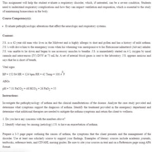
This assignment will help the student evaluate a respiratory disorder, which, if untreated, can be a severe condition. Students need to understand respiratory complications and how they can impact ventilation and respiration, which is essential to the study of maintaining homeostasis in the body.
Asthma Management
Course Competency(s):
- Evaluate pathophysiologic alterations that affect the neurologic and respiratory systems.
Content:
J.S. is a 42-year-old man who lives in the Midwest and is highly allergic to dust and pollen and has a history of mild asthma. J.S.’s wife drove him to the emergency room when his wheezing was unresponsive to his fluticasone/salmeterol (Advair) inhaler. J.S. was unable to lie down and began to use accessory muscles to breathe. J.S. is immediately started on 4 L oxygen by nasal cannula and intravenous (IV) D5W at 75 mL/hr. A set of arterial blood gases is sent to the laboratory. J.S. appears anxious and says that he is short of breath.
Vital signs
BP = 152/84 HR = 124 bpm RR = 42 Temp = 100.40F
ABGs
pH = 7.31 PaCO2 = 48 HCO3 = 26 PaO2 = 55
Instructions:
Investigate the pathophysiology of asthma and the clinical manifestations of the disease. Analyze the case study provided and determine what symptoms support the diagnosis of asthma. Identify the treatment provided in the emergency department and determine what additional therapies are needed to mitigate the asthma symptoms and return the client to wellness.
- Do you have any concerns with the numbers above?
- Identify what may be causing (aetiology) J.S. to have an exacerbation of asthma.
Prepare a 3-5 page paper outlining the causes of asthma, the symptoms that the client presents and the management of the disorder. Use at least one scholarly source to support your findings. Examples of literary sources include academic journals, textbooks, reference texts, and CINAHL nursing guides. Be sure to cite your sources in-text and on a References page using APA format.